


Fruit Fly Brain Mapping: A Leap Towards Understanding Human Neurology
Scientists map 50 million connections in fruit fly brain, advancing neuroscience. This milestone aids research on human brain disorders and complex behaviors, showcasing the power of tiny model organisms.
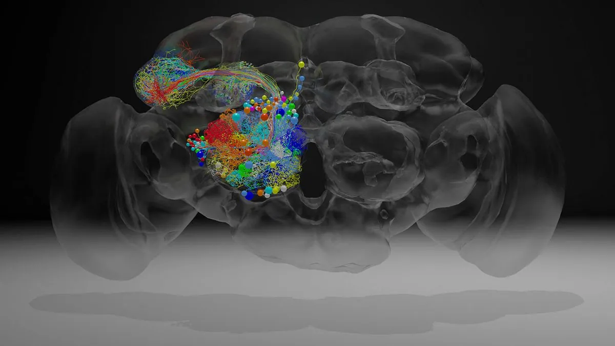
In a groundbreaking scientific achievement, researchers have successfully mapped over 50 million connections within the minuscule brain of a fruit fly. This intricate mapping, known as a connectome, represents the most detailed survey of an adult animal brain to date and marks a significant step towards understanding the complexities of the human brain.
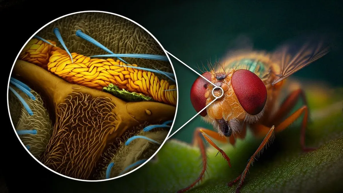
John Ngai, director of the National Institutes of Health's BRAIN Initiative, emphasizes the importance of this achievement. Despite their small size, fruit flies engage in a variety of complex behaviors, including eating, navigating towards smell signals, mating, and even exhibiting aggression. These tiny creatures, with brains about the size of a poppy seed, have been evolving for tens of millions of years and can perform sophisticated tasks like flying and perceiving ultraviolet light.
The BRAIN Initiative, launched in 2014 with an investment exceeding $3.5 billion, aims to enhance our understanding of the nervous system and accelerate the discovery of treatments for neurological disorders. This is particularly crucial given that over 40% of humans suffer from some form of nervous system disorder.
Fruit flies, along with other model organisms like zebrafish, roundworms, and mice, play a vital role in scientific research. These creatures share many genes with humans, with fruit flies possessing almost 75% of the genes involved in human diseases. This genetic similarity makes them invaluable for studying various aspects of biology and neuroscience.
The creation of this detailed brain map presented significant technical challenges. Davi Bock, co-senior author of one of the Nature papers and associate professor at the University of Vermont's Larner College of Medicine, describes the fruit fly brain as containing roughly 150 meters of wiring within a space smaller than a millimeter. The process involved removing the fly brain, turning it into a hard block, and cutting it into over 7,000 ultrathin sections using diamond knives.
These sections were then viewed under a high-speed electron microscope, resulting in 21 million images. The resolution achieved was incredibly fine, with each pixel representing a section of brain tissue about 4 by 4 by 40 nanometers. This level of detail was necessary to trace the intricate neural connections.
The project's success relied on collaboration between scientists and volunteers who helped proofread the data. State-of-the-art segmentation algorithms were used to convert the images into a form that allowed for the extraction of neuron chunks, which were then assembled into the final map.
This fruit fly connectome opens up numerous research possibilities. Scientists can now explore brain differences between male and female flies, adult and juvenile specimens, and healthy and diseased individuals. The map will aid in understanding how flies see, hear, sing, fly, and engage in complex behaviors like courtship and navigation.
Moreover, this research contributes to our understanding of how genes relate to brain function and influence brain wiring during development. The fruit fly model has already proven valuable in studying neurodegenerative diseases, the effects of drugs and alcohol on the nervous system, and even the genetic basis of obesity and longevity.
As we continue to unravel the mysteries of the fruit fly brain, we move closer to comprehending the intricacies of our own neural networks. This milestone in neuroscience, funded by the BRAIN Initiative and supported by the 21st Century Cure Act of 2016, paves the way for future breakthroughs in understanding and treating human neurological conditions.